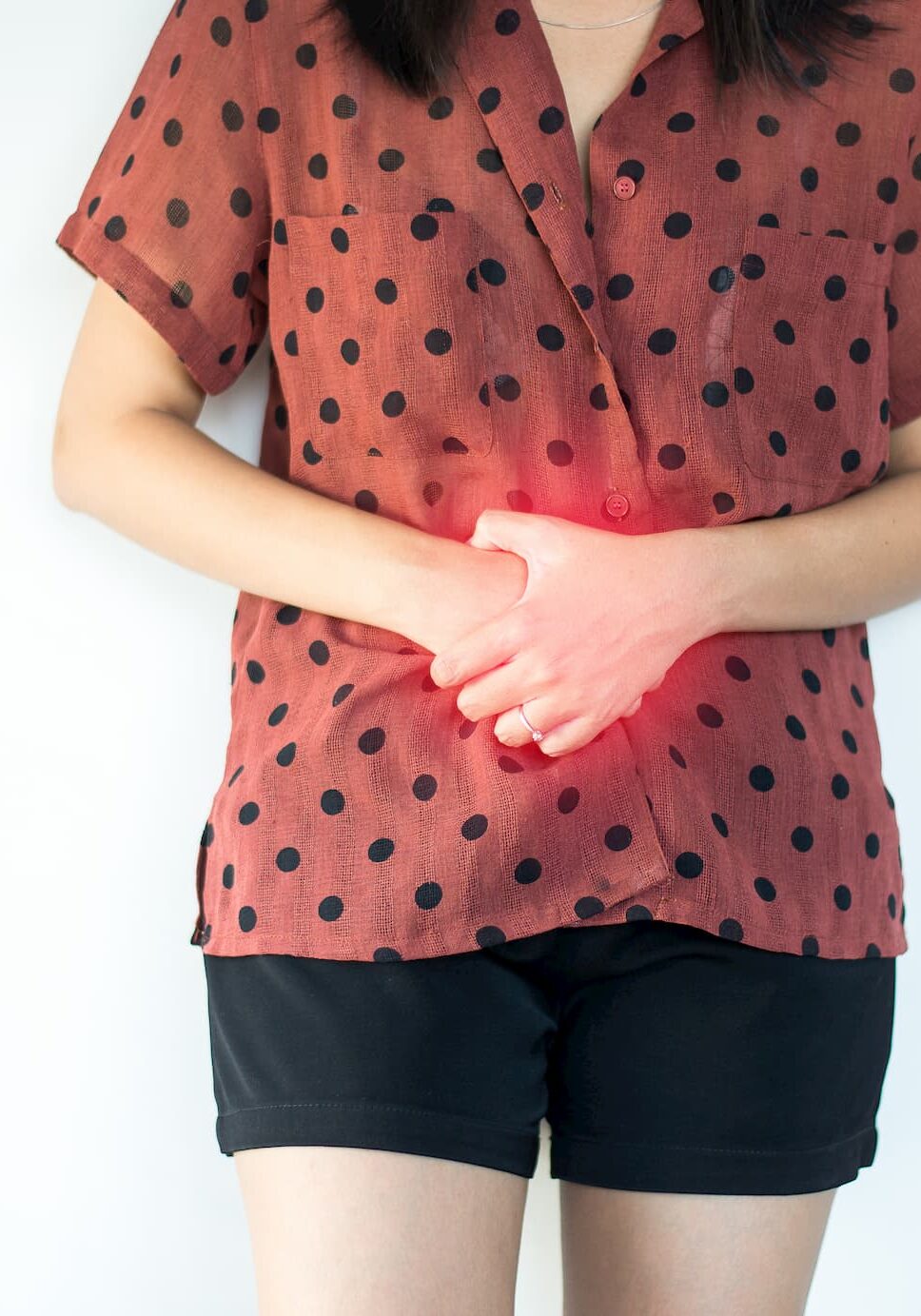
Gallstones are formed as crystals of cholesterol or pigments found in bile. They commonly occur in the gallbladder, where they can cause pain or lead to infection.
Gallstones can escape from the gallbladder where they can block the ducts causing jaundice or pancreatitis.
About a third of the population have gallstones. Women are three times as likely to develop gallstones compared to men. Although the incidence of gallstones increases with age, a younger age group is now being affected. Gallstones are more common in patients who are overweight and following dieting.
What are the symptoms / signs?
Most patients who have gallstones have no symptoms and do not know they have them. If you are found to have gallstones that are not causing symptoms, your doctor will discuss whether you ought to have them removed.
Stones can block the exit of the gallbladder, causing muscle spasm known as biliary colic. This generally lasts for one to two hours, often associated with feeling sick, normally requiring strong pain killers. Sufferers often describe the pain as coming on after eating, particularly after a fatty meal. Patients are best advised to avoid any foods that bring on the pain.
Infection within the gallbladder may follow. This is called “cholecystitis”. The pain is often severe enough to warrant admission to hospital for antibiotics and fluids.
Gallstones can escape into the bile ducts and cause problems. Smaller stones are more likely to cause problems than the larger solitary stones. Stones can become lodged in the bile duct, causing jaundice or even pancreatitis.
The pain caused by gallstones does not always follow the classic pattern and can often be misdiagnosed or mistaken for other problems.
Acid reflux or stomach ulcers can both present with sudden upper abdominal pain after eating. They may also be exacerbated by fatty foods. Both are common conditions, as are gallstones. They can both occur separately or together and often the only way to tell if the gallstones are the true cause of the pain, is to remove the patient’s gallbladder.

What investigations will be done?
Blood tests can help to confirm the presence of gallstones within the bile ducts by showing an effect on liver function.
Endoscopic Retrograde Cholangiopancreatography (ERCP)
Around 10% of people with gallstones will have stones in their bile ducts. If identified, these can be removed using ERCP. ERCP is a technique that combines the benefits of endoscopy with X-ray. The endoscope is a long video camera which is passed via your mouth, into your stomach and duodenum. If a stone is identified within the bile duct, the entrance can be widened with fine instruments and the stone removed.
An alternative is to remove bile duct stones at the same time as gallbladder removal surgery. This can usually be performed laparoscopically. Although bile duct surgery usually involves an extended recovery, it has the advantage of avoiding an additional hospital procedure.
What is the treatment?
The treatment of choice for gallstones is removal of the gallbladder - cholecystectomy.
Traditionally cholecystectomy was performed through a large cut in the right side of the abdomen. This produced a large wound that was painful, restricted breathing and required a long recovery period. Laparoscopic or keyhole surgery has now been universally adopted as the technique of choice for gallbladder removal. Under general anaesthetic, four small cuts are made in the patient’s abdominal wall (one at the naval and three below the ribs). A video camera is inserted through the naval to visualise the gallbladder. Long instruments are passed through the remaining holes to disconnect the gallbladder from the liver and bile ducts. The gallbladder can then be removed through one of the previously made holes. Around 2% of operations will have to be converted to an open operation, for reasons of safety. Reasons for this include excessive bleeding, very inflamed tissues or accidental damage to neighbouring structures. Previous upper abdominal surgery can increase the risk of conversion.
